
Radiology has played a pivotal role in the detection and diagnosis of aneurysms (localized dilatations of blood vessels) that can lead to life-threatening hemorrhage. Cutting-edge imaging modalities, such as CT angiography (CTA), magnetic resonance angiography (MRA), and digital subtraction angiography (DSA), offer noninvasive, high-resolution views of cerebral and abdominal vessels, empowering clinicians to detect aneurysms at early stages.
Advanced Imaging Techniques in Aneurysm Radiology
CT Angiography (CTA)
Often the first diagnostic tool utilized in aneurysm radiology is CT angiography (CTA), which rapidly records 2D and 3D images of blood arteries. CTA uses contrast dye to define vascular structures, thereby allowing radiologists to evaluate an aneurysm size, location, and form. For an aneurysm greater than 3 mm and for the detection of continuous bleeding, this approach is quite successful.
Magnetic Resonance Angiography (MRA)
Magnetic resonance angiography (MRA) offers excellent soft tissue contrast free from ionizing radiation. Methods such as time-of-flight (TOF) MRA and contrast-enhanced MRA especially benefit visualization of tiny and unruptured aneurysms. Furthermore, recent developments such as 7 T MRA push the boundaries of spatial resolution, which is essential for treatment planning.
Digital Subtraction Angiography (DSA)
Still the gold standard for vascular imaging is digital subtraction angiography (DSA). DSA provides extraordinary clarity and detail by removing pre-contrast images from post-contrast images; this is vital for preoperative planning in complicated aneurysm cases.
Integrating Radiology with Surgical Simulation
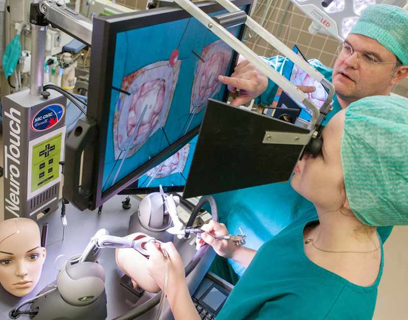
Modern diagnostic radiology not only facilitates accurate aneurysm detection but also enhances preoperative planning. Surgeons can use detailed radiological images to create patient-specific 3D models—an area where SurgeonsLab shines. SurgeonsLab’s advanced simulator models integrate real-time imaging data with virtual and physical simulation, allowing neurosurgeons and interventional radiologists to rehearse procedures in a risk-free environment. This special integration enhances patient outcomes and optimizes surgical approach.
The Future of Aneurysm Diagnosis and Training
In short, constant improvements in modeling and imaging technology are transforming aneurysm control. Machine learning models and deep learning frameworks are beginning to assist in automated aneurysm detection, ensuring that even subtle aneurysms are identified promptly. With radiology and simulation training merging seamlessly, the era of personalized, patient-specific preoperative planning is now here, making SurgeonsLab a leader in this innovative field.
SurgeonsLab’s simulator model, with its unparalleled realism and integration of advanced imaging, offers the only resource you'll ever need to master aneurysm radiology and treatment.


Write a comment ...